
THE STRUCTURE AND FUNCTION OF NEURONS

SPECIFICATION
THE STRUCTURE AND FUNCTION OF SENSORY, RELAY AND MOTOR NEURONS.
THE PROCESS OF SYNAPTIC TRANSMISSION: EXCITATION AND INHIBITION
KEYWORDS FOR NEURONS
ACTION POTENTIAL: An Impulse, or nerve impulse, is an action potential; it is an electrical signal that travels along a nerve channel. A nerve impulse occurs due to the difference in electrical charge across the plasma membrane of a neuron.
AXON: Axon is a tube-like structure that carries electrical impulses from the cell body to the axon terminals that pass the impulse to another neuron.
AXON-TERMINALS: Axon terminals (also called synaptic boutons, terminal boutons, or end-feet) are distal terminations of the telocentric (branches) of an axon.
AXON-HILLOCK: The axon hillock acts as something of a manager, summing the total inhibitory and excitatory signals. If the sum of these signals exceeds a threshold, the action potential is triggered, and an electrical signal is then transmitted down the axon away from the cell body. The axon hillock connects to the axon, an important structure that enables the propagation of the action potential, an electrical signal, down the axon.
CELL-BODY: Each neuron has a cell body with a nucleus, Golgi body, endoplasmic reticulum, mitochondria and other components.
DENDRITES: These are branch-like structures that receive messages from other neurons and allow the transmission of messages to the cell body.
EXCITATION & INHIBITION: A neurotransmitter is a signalling molecule secreted by a neuron to affect another cell across a synapse. The cell receiving the signal, any central body part or target cell, may be another neuron, but could also be a gland or muscle cell.
Mitochondria - produce energy to fuel cellular activities.
MYELIN-SHEATH: Myelin sheath is a substance which is found on neurons within the central nervous system (CNS) and the peripheral nervous system (PNS). The myelin sheath is the protective layer that wraps around the axons of neurons, insulating them and increasing the number of electrical signals transmitted.
NEURON: Neurons are the fundamental unit of the nervous system, specialised to transmit information to different body parts. “Neurons are the building blocks of the nervous system. They receive and transmit signals to different parts of the body. This is carried out in both physical and electrical forms. Several types of neurons facilitate information transmission. Sensory neurons transmit information from sensory receptor cells throughout the body to the brain. Motor neurons transmit information from the brain to the muscles. Interneurons transmit information between neurons in the body.
NEUROTRANSMITTER: A neurotransmitter is a signalling molecule secreted by a neuron to affect another cell across a synapse. The cell receiving the signal, any central body part or target cell, may be another neuron, but could also be a gland or muscle cell. Neurotransmitters are released from synaptic vesicles into the synaptic cleft, where they can interact with neurotransmitter receptors on the target cell. The neurotransmitter's effect on the target cell is determined by the receptor to which it binds. Neurotransmitters play a critical role in neural communication, influencing everything from involuntary movements to learning to mood. This system is both complex and highly interconnected. Neurotransmitters act in specific ways but can also be affected by diseases, drugs, or the actions of other chemical messengers.
NODES OF RANVIER. Gaps between the myelin sheath where the action potential jumps as it travels down the axon.
POST-SYNAPTIC CLEFT: Axon terminals where the neurotransmitters/signals are released from vesicles
PRE-SYNAPTIC CLEFT: Dendrites where neurotransmitters/signals are received.
RESTING POTENTIAL: A resting neuron −60 to −95 millivolts
ACTION POTENTIAL: An active neuron +30 millivolts. •
SYNAPSE: Synapse is also known as a neuronal junction, as it connects two neurons. They are the sites of transmission of electrical nerve impulses or chemical signals between neurons. It contains a small gap that separates neurons. It is the chemical junction between the terminal of one neuron and the dendrites of another neuron. Neurotransmitters are released from synaptic vesicles into the synaptic cleft, where they can interact with neurotransmitter receptors on the target cell. The receptor determines the neurotransmitter's effect on the target cell it binds.
VESICLES: Inside the axon terminal of a neuron are many synaptic vesicles. These are membrane-bound spheres filled with neurotransmitter molecules. There is a small gap between the axon terminal of the presynaptic neuron and the membrane of the postsynaptic cell, and this gap is called the synaptic cleft.
NEURON STRUCTURE: A neuron varies in shape and size depending on its function and location. All neurons have three parts: dendrites, a cell body, and an axon.
PARTS OF NEURON: The following are the different parts of a neuron:
SENSORY NEURONS: The sensory neurons convert signals from the external environment into corresponding internal stimuli. Sensory inputs activate sensory neurons and convey information to the brain and spinal cord. They are pseudo-unipolar in structure.
MOTOR NEURONS: These are multipolar and are located in the central nervous system, with their axons extending outside it. This is the most common type of neuron and transmits information from the brain to the body's muscles.
RELAY NEURONS: They are multipolar in structure. Their axons connect only to the nearby sensory and motor neurons. They facilitate the transmission of signals between neurons.
CHEMICAL SYNAPSE: In chemical synapses, the action potential affects other neurons through a gap between the two neurons, known as the synapse. The action potential is conducted along the axon to a postsynaptic terminal, where it initiates the release of neurotransmitters. These neurotransmitters excite postsynaptic neurons, which generate action potentials of their own.
ELECTRICAL SYNAPSE: A gap junction that connects two neurons forms an electrical synapse. These gaps include ion channels that help in the direct transmission of a positive electrical signal. These are much faster than chemical synapses.
WHAT IS A NEURON?
Consciousness, mobility and sensory perception are all the result of brain activity. The brain is composed of glial cells, including astrocytes. Among these cells are neurons – specialised cells whose function is to communicate and process information from the environment to the brain and vice versa. It does this by enabling neurons to communicate with one another via electrical and chemical signals. The human body is made up of trillions of cells. The human brain contains approximately 100 billion neurons, and each neuron is connected to approximately 1000 other neurons. This creates highly complex neural networks that confer the brain's impressive processing capabilities. Neurons are essential components of a vast communication system within the body.
Neurons are the oldest and longest cells in the body! You have many of the same neurons for your entire existence. Although other cells are replaced, many neurons are not. In fact, you have fewer neurons when you are old than when you are young. Neurons can be pretty large - neurons that control the spine can be several feet long! For communication between neurons to occur, an electrical impulse must trigger the release of chemicals. These chemicals, called neurotransmitters, allow other neurons in a circuit to be switched on or off. Ultimately, neurons communicating with each other are why humans can walk, talk, see, think, laugh, get angry, breathe, and sleep (amongst many other things).
Neurons are specialised cells that carry neural information throughout the body. Neurons can be one of three types: sensory, relay, or motor. Neurons typically consist of a cell body, dendrites and an axon. Dendrites at one end of the neuron receive signals from other neurons or sensory receptors. Dendrites are connected to the cell body, the neuron's control centre. From the cell body, the impulse is carried along the axon, where it terminates at the axon terminal. In many nerves, including those in the brain and spinal cord, there is an insulating layer that forms around the axon – the myelin sheath. This allows nerve impulses to transmit more rapidly along the axon. If the myelin sheath is damaged, conduction slows. The length of a neuron can vary from a few millimetres to one metre.
SENSORY, RELAY AND MOTOR NEURONS
There are three main types of neurons: sensory, relay, and motor. Each of these neurons has a different function, depending on its location in the body and its role within the nervous system. Note: All three types of neurons consist of similar parts. However, their structure, location, and function differ somewhat, and you should be aware of this.
Sensory neurons are found in receptors such as the eyes, ears, tongue and skin, and carry nerve impulses to the spinal cord and brain. When these nerve impulses reach the brain, they are translated into ‘sensations’, such as vision, hearing, taste and touch. However, not all sensory neurons reach the brain; some terminate in the spinal cord, enabling rapid reflex responses.
Relay neurons are found between sensory input and motor output/response. Relay neurons are found in the brain and spinal cord and allow sensory and motor neurons to communicate.
Motor neurons are found in the central nervous system (CNS) and control muscle movements. When motor neurons are stimulated, they release neurotransmitters that bind to receptors on muscle cells, triggering a response that leads to movement.
As you can see from the diagrams above, all three neurons consist of similar parts. The dendrites receive signals from other neurons or sensory receptor cells. The dendrites are typically connected to the cell body, which is often referred to as the ‘control centre’ of the neuron, as it contains the nucleus. The axon is a long, slender fibre that carries nerve impulses, known as action potentials, away from the cell body towards the axon terminals, where the neuron ends. Most axons are surrounded by a myelin sheath (except for relay neurons), which insulates the axon so that the electrical impulses travel faster along the axon. The axon terminal connects the neuron to other neurons (or directly to organs) via synaptic transmission.
HOW DOES NEURAL TRANSMISSION WORK?
All things need energy to work
Cars require petrol, plants require sunlight, animals require food, and TVs require electricity. Neurons are no different; if they are to fire neurotransmitters (chemicals) at other neurons and transmit messages, some form of energy must power their function.
LAYMAN VERSION OF NEURAL TRANSMISSION
In essence, the neuron runs on electricity; it is the neuron's battery or power source. What triggers the electricity to fire neurotransmitters is strangely enough, other neurotransmitters, It is a never-ending cycle of neurotransmitters landing on the head of a neutron (dendrites), causing electrical spurge that travels down the neutron’s body (axon) that makes the tail of the neuron (axon terminals) release more neurotransmitters that float onto the head’s (dendrites of other neurons and effectively switch them on by causing them to send electricity (action potential) down the neutron and so on and on. In other words, brain chemistry.
SCIENTIFIC VERSION OF NEURAL TRANSMISSION
Neurotransmitters are received on the dendrites of a neutron these neurotransmitters will then be summed up at the top of the axon in a part called the axon Hillock if an insufficient amount of neurotransmitters are received on the Hillock they will not trigger an action potential (an bit like putting the wrong amount of AA batteries in a torch, if you put two batteries in the torch instead of 3, then the torch will not turn on..but if an adequate amount signals are built up at the axon hillock, then an action potential is triggered which sends electricity down the axon. When the electricity reaches the end of the axon, it travels to the axon terminals (also called the presynaptic terminals), where it will trigger the vesicles (holding tanks containing neurotransmitters) to release their contents into the synaptic cleft toward the dendrites (postsynaptic membranes) of other neurons in that connection/network.
SYNAPSES
Once an action potential arrives at the terminal button at the end of the axon, it must be transmitted to another neuron or to tissue/muscle. To achieve this, it must cross the synaptic cleft between the presynaptic and postsynaptic neurons. This area is known as the synapse. The physical gap between the presynaptic and postsynaptic cell membranes is known as the synaptic gap. At the end of the axon of the nerve cell are several sacs known as synaptic vesicles. These vesicles contain chemical messengers (neurotransmitters, chemicals in the brain). As the action potential (electrical signal) reaches the synaptic vesicles, it triggers the release of their contents. The released neurotransmitter (such as serotonin or dopamine) diffuses across the gap between the pre-and postsynaptic cells, where it binds perfectly to specialised receptors that recognise it (a bit like a lock and key) and that are activated by that particular neurotransmitter.
Once the neurotransmitter crosses the gap and has been taken up by the post-synaptic receptor site, i.e. the dendrites of the next neuron, the chemical message is converted back into an electrical impulse, and the transmission process begins again in this other neuron. A single axon can have multiple branches, allowing it to make synapses on various postsynaptic cells. Similarly, a single neuron can receive thousands of synaptic inputs from many different presynaptic neurons. Until recently, it was thought that a neuron produced and released only one type of neurotransmitter. This was called "Dale's Law." However, there is no evidence that neurons can contain and release more than one type of neurotransmitter.
EXCITATION AND INHIBITION OF NEURONS
SCENARIO ONE
Imagine that you’re driving down a road undeterred, with no red lights or stop signs to slow you down. While that may seem like a fascinating idea, it is obviously hazardous since our roads are not all parallel but interconnected in several different ways. To ensure smooth traffic flow in all directions, we use stop signs, red lights, speed bumps, and police patrols to prevent accidents. In much the same way, our brain has a mechanism to keep the excitation in check. Information in the brain flows via excitatory neurons whose properties depend on their anatomical location. For example, a neuron in the visual cortex will respond to visual stimuli, and a neuron in the auditory cortex will respond to auditory stimuli. Since excitation cannot persist indefinitely, we must ensure that it decays or ceases whenever required. This is known as inhibition. Inhibition is as essential as excitation, if not more so. The neurons that perform this function are known as inhibitory neurons, and they have the special property of ensuring that the brain functions smoothly and is accident-free.
When activated, inhibitory neurons release the neurotransmitter GABA, which hyperpolarises the postsynaptic neuron; i.e., it makes the membrane potential more negative, making it harder for the neuron to reach the threshold to fire an action potential, thereby causing ‘inhibition’. Most often, inhibitory neurons are also called GABAergic neurons for that reason. Although they constitute only 20-25% of all neurons in the cortex, they are strikingly diverse, with different morphologies, sizes, intrinsic properties, connectivity patterns, and protein expression. Based on their molecular properties, a significant effort
EXCITATORY AND INHIBITORY NEUROTRANSMITTERS
Neurotransmitters can be classified as excitatory or inhibitory, exerting one of these two effects on the neighbouring neuron. For instance, the neurotransmitter serotonin induces inhibition in the receiving neuron, thereby making the neuron more negatively charged and less likely to fire. Inhibitory neurotransmitters are like the nervous system’s “off switches” and are generally responsible for calming the mind and body, inducing sleep, and filtering out unnecessary excitatory signals. An inhibitory neurotransmitter binding with a postsynaptic receptor results in an inhibitory postsynaptic potential (IPSP), making it less likely to fire.
In contrast, neurotransmitters such as noradrenaline are excitatory; they are the nervous system’s “on switches”. These cause postsynaptic neuron excitation by increasing its positive charge and making it more likely to fire. It induces an electrical charge across the cell membrane, resulting in an excitatory postsynaptic potential (EPSP) and increasing the likelihood of firing. A nerve cell can receive both EPSPs and IPSPs at the same time. The likelihood of the cell firing is determined by summing the excitatory and inhibitory synaptic inputs. The net sum of this calculation (summation) determines whether or not the cell fires.
The nervous system controls the body’s organs and plays a role in nearly all bodily functions. Nerve cells, also known as neurons, and their neurotransmitters play essential roles in this system. Neurons fire impulses. They release neurotransmitters, also known as the body’s chemical messengers. These chemicals carry signals to other cells.
WHY NEURONS CAN ONLY TRANSMIT INFORMATION IN ONE DIRECTION AT A SYNAPSE.
A Nerve electrical impulse only travels in one direction. There are several reasons why nerve impulses travel only in one direction. The most important is synaptic transport. For a "nerve impulse" to pass from cell to cell, it must cross synaptic junctions. Nerve cells are aligned head-to-tail along a nerve tract and are not connected; they have tiny gaps between them and the next cell. These tiny gaps are called synapses. When you get a nerve firing, you have probably heard that it is an electrical impulse that carries the signal. This is true, but it is not electrical, unlike your wall outlet. This is electrochemical energy. Neurotransmitters are molecules that fit like a lock and key into a specific receptor. The receptor is located on the next cell in the line. When the neurotransmitter hits the receptor on the next cell in line, it signals that cell to begin firing as well.
This will continue along the entire length of the nerve tract. In brief, a nerve impulse results in a chain reaction along the nerve cell's axon, or stemlike section. Sodium (Na+) ions flow in, potassium (K+) ions flow out, and an electrochemical gradient develops along the length of the cell. You can think of it as a line of gunpowder that someone lit, with the flame travelling down the length of it. Common electrical power is more like a hose full of water, and when you apply pressure at one end, the water jets out the other. Therefore, nerve impulses cannot travel in the opposite direction because nerve cells have only neurotransmitter storage vesicles that travel one way and receptors in one place.
WHAT ARE NEUROTRANSMITTERS?
Neurotransmitters are chemical messengers that your body can't function without. Their role is to carry chemical signals (messages) from one neuron to the next. The next target may be another neuron, a muscle, or a gland. Each neurotransmitter attaches to a different receptor. For example, dopamine molecules attach to dopamine receptors. When they attach, it triggers an action in the target cells.
WHAT DO NEUROTRANSMITTERS DO?
The brain needs neurotransmitters to regulate many necessary functions, including:
Heart rate
The fight or flight response
Breathing
Sleep cycle wake cycle
Digestion
Mood
Concentration
Regulating appetite
Muscle movement
Memory
Low levels of any neurotransmitter can lead to problems, including fibromyalgia, Parkinson’s disease and Alzheimer's disease. Imbalances can also cause psychiatric conditions such as anxiety, depression, schizophrenia, and violence. Over 100 neurotransmitters have been identified and are still being identified, but only seven do most of the work. These seven neurotransmitters are acetylcholine, dopamine, gamma-aminobutyric acid (GABA), glutamate, histamine, norepinephrine, and serotonin. After neurotransmitters deliver their messages, the body degrades or recycles them.
TYPES OF NEUROTRANSMITTERS
Neurotransmitters have different types of actions:
Excitatory neurotransmitters encourage a target cell to take action.
Inhibitory neurotransmitters reduce the likelihood that the target cell will take action. In some cases, these neurotransmitters have a relaxation-like effect.
Modulatory neurotransmitters can send messages to many neurons at the same time. They also communicate with other neurotransmitters.
Some neurotransmitters can perform multiple functions depending on the type of receptor to which they bind.
NEUROTRANSMITTERS AND THEIR FUNCTIONS
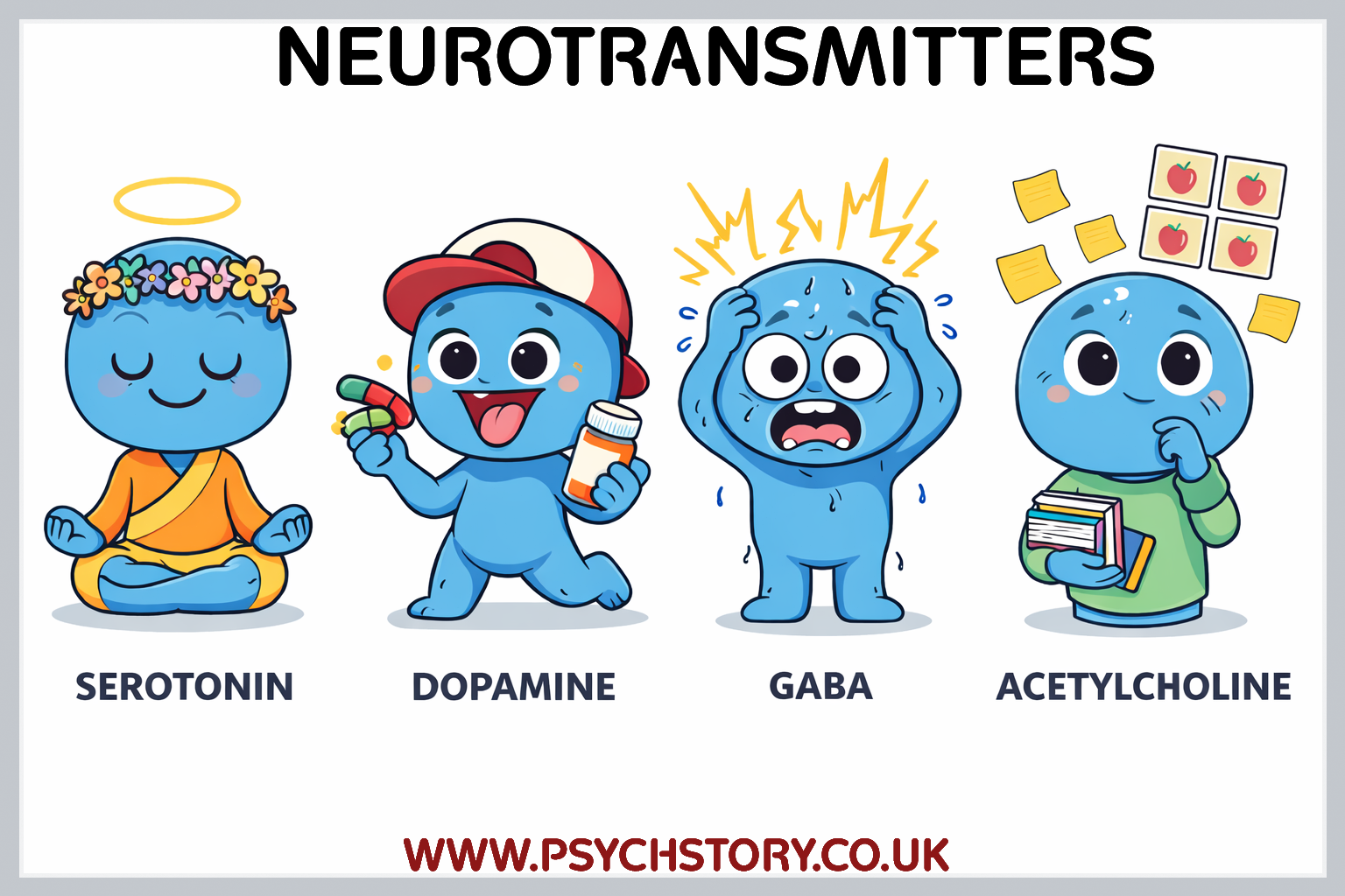
ACETYLCHOLINE (EXCITATORY)
PRIMARY FUNCTION: Muscle contraction, attention, arousal, learning and memory.
OPTIMUM LEVELS: Focus, attention, memory, learning, muscle control.
EXCESS: —
DEFICIENCY: Alzheimer’s disease, fatigue, inattention, ADD/ADHD.
DRUGS THAT ACT ON IT: Donepezil (cholinesterase inhibitor), Nicotine (agonist), Botulinum toxin (antagonist).
DOPAMINE (MAINLY EXCITATORY)
PRIMARY FUNCTION: Motivation, reward, pleasure, focus, voluntary movement, attention and learning.
OPTIMUM LEVELS: Motivation, pleasure, focus, reward-seeking behaviour.
EXCESS: Hallucinations, delusions, paranoia, mania.
DEFICIENCIES: Parkinson’s disease, anhedonia, lack of motivation, ADHD, depression.
DRUGS THAT ACT ON IT: L-DOPA (precursor), Antipsychotics (D2 receptor antagonists), Cocaine & Amphetamines (increase release), Methylphenidate (reuptake inhibitor).
ENKEPHALINS AND ENDORPHINS (INHIBITORY)
PRIMARY FUNCTION: Natural pain relief, pleasure, stress reduction, euphoria.
OPTIMUM LEVELS: Pain relief, pleasure, stress reduction.
EXCESS: Can be caused by opioid drugs or certain medical conditions. Leads to excessive sedation and respiratory depression
DEFICIENCY: Increased pain, sensitivity.
DRUGS THAT ACT ON IT: Opioid agonists (Heroin, Morphine, Codeine, Oxycodone, Fentanyl, Methadone).
ADRENALINE (EPINEPHRINE) (EXCITATORY)
PRIMARY FUNCTION: Fight-or-flight response, arousal, energy, quick decision-making.
OPTIMUM LEVELS: Arousal, energy, drive, excitement.
EXCESS: Anxiety, high blood pressure, chronic stress.
DEFICIENCY: Fatigue, lack of drive, depression.
DRUGS THAT ACT ON IT: Epinephrine (EpiPen), some sympathomimetic drugs.
NORADRENALINE (NOREPINEPHRINE) (EXCITATORY)
PRIMARY FUNCTION: Fight-or-flight response, alertness, memory formation, metabolic rate.
OPTIMUM LEVELS: Alertness, focus, energy.
EXCESS: Anxiety, agitation, high blood pressure.
DEFICIENCY: Depression, low energy, poor concentration.
DRUGS THAT ACT ON IT: SNRIs (e.g. Venlafaxine), Atomoxetine.
GABA (INHIBITORY)
PRIMARY FUNCTION: Primary inhibitory neurotransmitter – reduces neuronal excitability.
OPTIMUM LEVELS: Calmness, reduced anxiety, stable mood.
EXCESS: Excessive sedation, memory impairment.
DEFICIENCY: Anxiety, insomnia, epilepsy, schizophrenia.
DRUGS THAT ACT ON IT: Benzodiazepines (GABA-A receptor agonists), Alcohol, Barbiturates.
SEROTONIN (MAINLY INHIBITORY)
PRIMARY FUNCTION: Mood regulation, sleep, appetite, and emotional stability.
OPTIMUM LEVELS: Contentment, good mood, serenity, stable emotions.
EXCESS: Serotonin syndrome.
DEFICIENCY: Depression, anxiety, OCD, insomnia, aggression.
DRUGS THAT ACT ON IT: SSRIs (selective serotonin reuptake inhibitors), MDMA (serotonin releaser).
SUBSTANCE P (EXCITATORY)
PRIMARY FUNCTION: Transmission and perception of pain.
OPTIMUM LEVELS: Normal pain signalling.
EXCESS: Often linked to inflammation, injury, chronic pain conditions, or certain medications. Leads to increased pain sensitivity.
DEFICIENCY: Reduced pain perception.
DRUGS THAT ACT ON IT: Capsaicin (depletes Substance P).
QUESTIONS ON THE STRUCTURE AND FUNCTION OF SENSORY, RELAY AND MOTOR NEURONS. AND THE PROCESS OF SYNAPTIC TRANSMISSION: EXCITATION AND INHIBITION
Label a neuron: synapse, myelin-sheath, nodes of Ranvier, axon-hillock, pre-synaptic cleft, post-synaptic cleft, vesicles, cell-body, dendrites, axon, axon-terminals.
What is the chemical released into the synaptic gap called? Neurotransmitter
What is an action potential? Please draw an arrow above the neuron indicating.
What direction should the action potential travel down the axon?
What is the action potential of a neuron in milliseconds?
What is the axon hillock?
What role does the axon hillock have in an action potential?
Explain why neurons can only transmit action potentials in one direction.
What is a:
Sensory neuron
Motor neuron
Relay neuron
Where are the three types of neurons located?
Information can only travel in one direction at a synapse. Explain why neurons can only transmit information in one direction at a synapse.
Answer: Because of the chemical nature of impulse and the axon-dendrite structure.