EARLY NERVOUS SYSTEMS: FROM NERVE NET TO NEOCORTEX
EARLY NERVOUS SYSTEMS: FROM NERVE NET TO NEOCORTEX
RECAP AND ASSESSMENT
Before starting Localisation of Function, make sure you’re confident with the basics of brain structure and terminology. If you need a quick refresher on the central regions of the brain and what they do, visit BRAIN ANATOMY AND FUNCTION.
Once you’re ready to dive deeper, you can explore how this topic is assessed — including essay guidance, sample responses, and examiner-style tips — in ASSESSMENT MATERIALS FOR LOCALISATION OF FUNCTION.
SAME FOUNDATIONS, BUT WILDLY DIFFERENT FOUNDTIONS
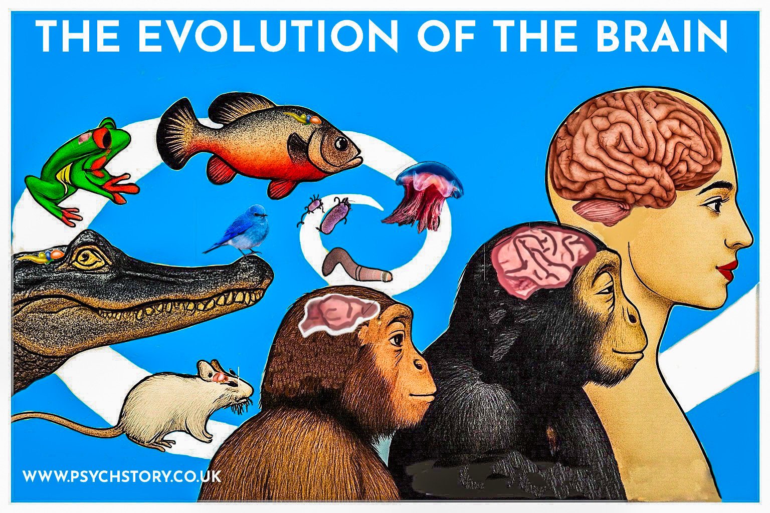
Understanding how the neocortex is organised begins with seeing how it was built. The brain’s structure did not appear fully formed; it evolved step by step, with new parts added to old ones and each taking on specialised jobs. Tracing that journey — from the first nerve nets in simple animals to the layered neocortex of mammals — shows why the modern cortex is arranged the way it is. It explains how different regions came to perform distinct functions and why the human brain, though complex, still follows the same basic plan as our earliest ancestors.
Early nervous systems began with a nerve net, the brain's basic foundation, which, over time, diverged and adapted to different environmental pressures. The earliest brains, such as the nerve net of a jellyfish, consist of a diffuse web of neurons capable only of simple reflexes: pulsing to swim, stinging in response to touch, and regulating heartbeat. There is no capacity for learning, memory, or planning — only immediate, automatic survival.
Over hundreds of millions of years, as environments became more complex and demanding, the early bilaterian nervous system was conserved and elaborated. Evolution added new layers of organisation, connecting and refining existing circuits rather than replacing them. Each adaptation enhanced perception, coordination, memory, or behavioural flexibility while maintaining continuity with the primitive central nervous system that first enabled integrated sensation and movement.
A good analogy for this is a bungalow house — a simple one-storey building with four walls and a roof that fulfils only the most essential needs. This basic structure provides the foundation for more complex dwellings. However elaborate those later designs become, they all retain the same core elements: walls, ceilings, and a central framework.
In the same way, every nervous system retains this ancestral architecture.
EVOLUTION SOLUTIONS
The human brain did not appear all at once. It developed through many small changes over millions of years as animals adapted to different environments. There was no single path — evolution tried many approaches. Some species changed in separate directions, others found the same solutions in different ways, and some even evolved together. In humans, culture itself became part of evolution, feeding back into the brain’s growth. These main patterns — divergent, convergent, co-evolution, and gene–culture coevolution — show how complex brains can arise from simple beginnings.
DIVERGENT EVOLUTION happens when a species that shares a common ancestor evolves along different paths as it adapts to new environments. For example, the forelimbs of mammals all share a basic bone structure but have been reshaped for different purposes: wings in bats, flippers in whales, and grasping hands in primates. The exact process happens in the brain: species start with the same neural plan but emphasise different regions. A mole’s brain, for instance, devotes more area to touch, while an owl’s gives greater space to vision and hearing.
COVERGENT EVOLUTION happens when unrelated species face similar environmental demands and evolve similar solutions. Birds and bats, for example, both developed the ability to fly, even though their ancestors were very different. Likewise, dolphins and sharks both evolved streamlined bodies for swimming, even though one is a mammal and the other a fish. In the same way, dolphins, elephants, and humans have each developed large, folded brains capable of complex communication and problem-solving — not because they share a recent common ancestor, but because social living and cooperation favour intelligence.
In CO-EVOLUTION, two species influence each other’s development over time. One's improvements create new challenges for the other. A classic example is the evolutionary “arms race” between bats and moths: bats evolved echolocation to hunt, and some moths evolved ears that detect ultrasonic calls, allowing them to dodge attacks. Each side drives the other’s sensory and neural adaptations.
Finally, GENE-CULTURE COEVOLUTION: THE HUMAN FEEDBACK LOOP, is found only in humans, describes how culture and biology mutually shape one another. When humans began farming, for example, people who could digest milk as adults had a strong nutritional advantage, so genes for lactase persistence spread quickly. Similarly, the ability to use language and tools created new pressures that favoured brains capable of planning, memory, and communication. Over time, these cultural practices and genetic changes reinforced one another, leading to the enormous and flexible human brain.
The human brain is the endpoint of a long architectural project. Each evolutionary step built upon an earlier design, adding new control systems, sensory maps, and layers of processing without ever discarding the old ones. To understand how this ancestral framework evolved into the modern cortex, it helps to trace the major milestones in nervous system evolution—from the earliest diffuse networks of nerve cells to the highly folded cerebral hemispheres of humans.
THE ROAD TO THE NEOCORTEX
The neocortex is unique to mammals. It has six layers of neurons, stacked and interconnected vertically to form columns of processing cells. These columns act like tiny circuits that handle one small piece of information at a time — such as the edge of an object, the direction of a sound, or the position of a limb — before combining that information into a complete perception.
The evolution of the nervous system reflects a steady increase in complexity, connectivity, and specialisation. Each stage adds new structures and refinements while retaining the original plan.
HOW THE NEOCORTEX EVOLVED
The cortex emerged gradually from older brain tissue called the pallium, which covered the forebrain in early vertebrates such as fish and amphibians. The pallium could process sensory information, but in a simple, unlayered way. As evolution advanced, this tissue began to divide into zones with more specialised roles. By the time mammals appeared, these zones had become distinct regions:
ARCHICORTEX — the oldest region, seen today in the hippocampus, is involved in memory and navigation.
PALEOCORTEX — a slightly newer region linked to smell and emotion.
NEOCORTEX — the newest and largest region, responsible for flexible thought, sensory perception, and reasoning.
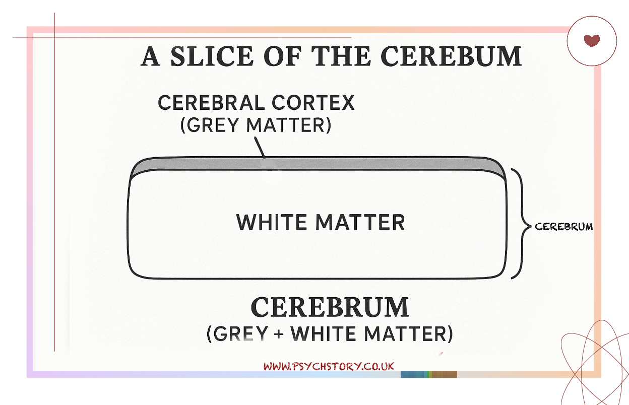
Together, these regions form the cerebral cortex — the sheet of outer grey matter that integrates sensation, memory, and action. The neocortex is the most recently evolved and most elaborate part of the brain. The cortex is not a new invention but a refined extension of ancient neural structures.
To see how this architecture took shape, we can trace the key milestones in nervous system evolution, from the simplest nerve nets to the layered forebrains of mammals.
THE FIRST BLUEPRINT
Early animals, such as jellyfish, had only a nerve net — a loose weave of nerve cells that coordinated pulsing and movement but lacked a central control centre.
Example species: jellyfish, sea anemone, hydra.
THE FIRST COORDINATION SYSTEM
Flatworms and simple chordates acquired a spinal cord, a central line of communication connecting the ends of the body. This was the first wiring trunk through which information could travel quickly.
Example species: planarian worm, amphioxus (lancelet).
THE BRAINSTEM: AUTOMATIC CONTROL
As vertebrates evolved, part of the spinal cord enlarged to form the brainstem. This became the body’s life support system, managing heartbeat, breathing, and basic reflexes — functions that must never fail but do not require thought.
Example species: lamprey, hagfish.
THE HINDBRAIN: MOVEMENT AND BALANCE
The next addition was the cerebellum, a structure that fine-tuned motion and posture. Fish used it to swim efficiently; later species used it for walking and grasping.
Example species: bony fish, amphibians.
THE FOREBRAIN: SENSING AND LEARNING
The forebrain grew to process incoming sensory information, such as sight, smell, and touch and link it with memory and learning. It created the first flexible behaviour, the ability to change based on experience.
Example species: amphibians, early reptiles, birds.
THE CEREBRUM: DECISION MAKING
In reptiles and mammals, the forebrain expanded into the cerebrum, the large dome of tissue that makes up most of the human brain. Here, information from the senses is compared, decisions are made, and voluntary movements are initiated.
Example species: lizard, mouse, human
THE EMERGENCE OF THE CEREBRAL CORTEX
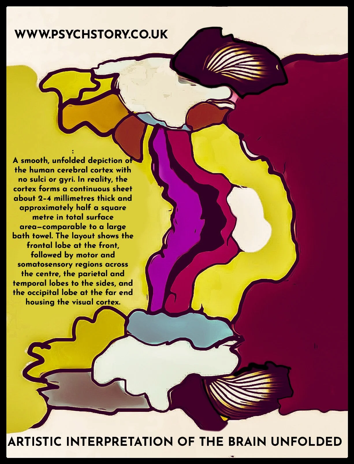
In mammals, this layer looks like a thin sheet of grey tissue covering the brain’s surface, about two to three millimetres thick. It is the most familiar part of the brain — the wrinkled surface seen in diagrams and models — and it enables complex, flexible behaviour.
When we talk about the cortex, we mean the outer layer. The word comes from the Latin corticis, meaning “bark,” because it covers the organ beneath it, just as bark covers a tree. Many organs have cortices: the adrenal cortex on the adrenal glands produces hormones, and the renal cortex on the kidneys filters blood. So, “cortex” by itself means outer covering.
The cerebral cortex, however, refers specifically to the outer layer of the cerebrum, the most significant part of the brain. It is made mainly of grey matter — the cell bodies and dendrites of billions of neurons responsible for processing, integrating, and generating information. It looks like a thin sheet of grey tissue covering the brain’s surface, about two to three millimetres thick. It is the most familiar part of the brain — the wrinkled surface seen in diagrams and models — and it enables complex, flexible behaviour.
Beneath it lies white matter, formed by bundles of myelinated axons that link different brain areas, allowing rapid communication between them
THE NEOCORTEX

The cerebral cortex is responsible for perception, memory, thought, and voluntary movement. It is made of grey matter — the cell bodies and dendrites of billions of neurons that process and integrate information — supported by white matter beneath, which carries messages between cortical regions. In all mammals, the cortex serves as the command centre for complex behaviour, transforming sensory input into organised experience and purposeful action.
As evolution advanced, the cortex expanded and grew more specialised. Early mammals had small, smooth cortices sufficient for basic sensory processing and movement. But as environmental and social demands increased — the need to hunt strategically, communicate, care for offspring, or navigate group living — brain capacity had to expand. This created a structural problem: the skull could not enlarge indefinitely without making birth impossible.
As mammalian brains expanded, physical limits emerged. The skull could not enlarge indefinitely without compromising movement, balance, or the ability to be born. To increase processing capacity without increasing head size, the cortex folded in on itself, forming ridges (gyri) and grooves (sulci). This folding expanded the surface area and allowed far more neurons to fit within the same cranial space. The more folded the cortex, the greater its capacity for learning, sensory integration, and behavioural flexibility. It is like fitting a king-size bedsheet into a handbag — a crinkled mass.
In humans, this constraint became particularly acute. Bipedalism narrowed the pelvis and reduced the width available for childbirth, while the growing brain demanded more space. The only evolutionary compromise was to increase cortical surface area through further folding. Human infants are therefore born with large but still unfinished brains, which continue to grow and form connections long after birth. This trade-off between locomotion, reproduction, and brain size shaped both our anatomy and our extended period of childhood development — a biological investment in learning and intelligence.
Less intelligent mammals, such as shrews, have smooth, relatively simple cortices suited to instinctive behaviour. Mid-level mammals — cats, dogs, and hoofed animals — exhibit moderate folding, which supports more flexible learning and complex sensory integration. In highly social or intelligent mammals such as dolphins, elephants, whales, and great apes, the cortex expands dramatically. It folds into deep, intricate patterns, producing brains capable of planning, empathy, memory, and cooperation. These species also show high encephalisation quotients — brain size relative to body size — reflecting advanced cognition and social awareness.
From this same circuitry emerge abstraction and symbolism. The neocortex can detach thought from the immediate present, compare possibilities, and construct inner worlds of art, mathematics, morality, and belief. It is the source of narrative identity — the sense of a continuous “self” that remembers, plans, and interprets experience.
Earlier animals possessed forebrains that guided behaviour, but the mammalian neocortex added the power to imagine what does not yet exist, to reason beyond instinct, and to reflect on the fact of being conscious at all.
In humans, cortical expansion reached its peak. The prefrontal cortex became dominant, supporting foresight, language, moral reasoning, and abstract thought. Specialised neurons such as Von Economo cells emerged, enhancing rapid social perception and emotional intelligence. Together, these adaptations produced the uniquely flexible, self-reflective human mind.
SUMMARY
EARLY NERVOUS SYSTEMS: FROM NERVE NET TO NEOCORTEX
The brain evolved gradually, adding new structures to old ones over hundreds of millions of years.
The earliest animals, like jellyfish, had only a nerve net for simple reflexes and movement.
Flatworms developed the first spinal cords; vertebrates later added brainstems, cerebella, and forebrains for coordination and learning.
The cerebral cortex evolved from the pallium, an early sensory layer in fish and amphibians.
It is divided into three central regions:
ARCHICORTEX (hippocampus): memory and navigation.
PALEOCORTEX: smell and emotion.
NEOCORTEX: higher cognition, reasoning, and flexible thought.
Only mammals have a true neocortex, a six-layered structure of neuronal columns that processes sensory information and integrates perception.
As mammals evolved, the cortex expanded and folded (gyri and sulci) to fit more neurons into limited skull space — like folding a king-size bedsheet into a handbag.
Small mammals (e.g. shrews) have smooth cortices; mid-level mammals (cats, dogs, hoofed animals) show moderate folding; highly social mammals (dolphins, elephants, primates) have deeply folded, complex cortices.
In humans, folding reached its maximum due to bipedalism and restricted childbirth size, leading to larger but still-developing infant brains.
The expanded prefrontal cortex enabled language, planning, morality, and abstract reasoning.
From this same circuitry emerged self-awareness, imagination, and symbolic thought — the ability to think beyond instinct and reflect on one’s own mind.
Here is the link to the BBC Brain Story Documentary with Dr Susan Greenfield, Episode One: "All in the Mind":
Watch BBC Brain Story - Episode 1: "All in the Mind"
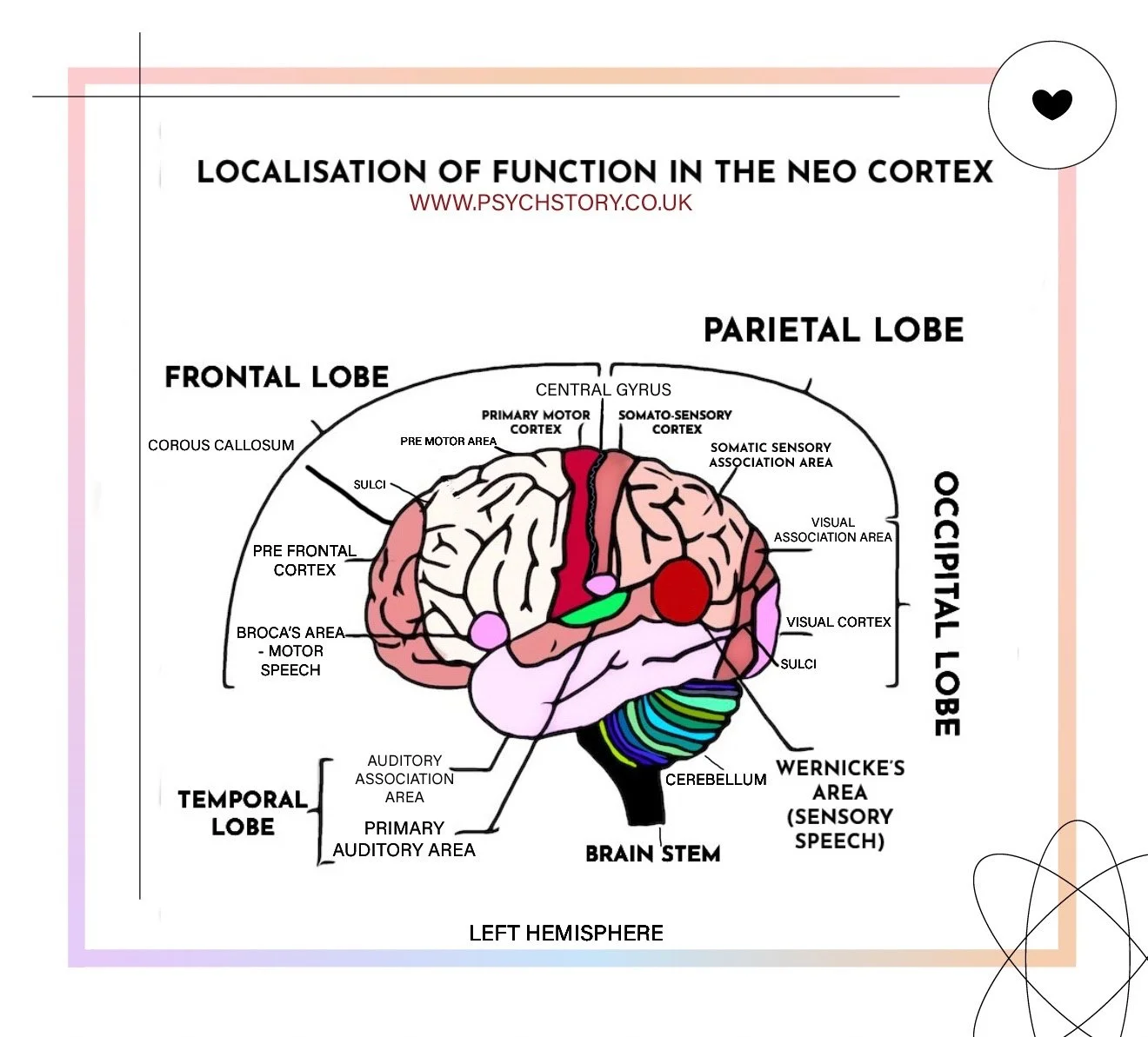
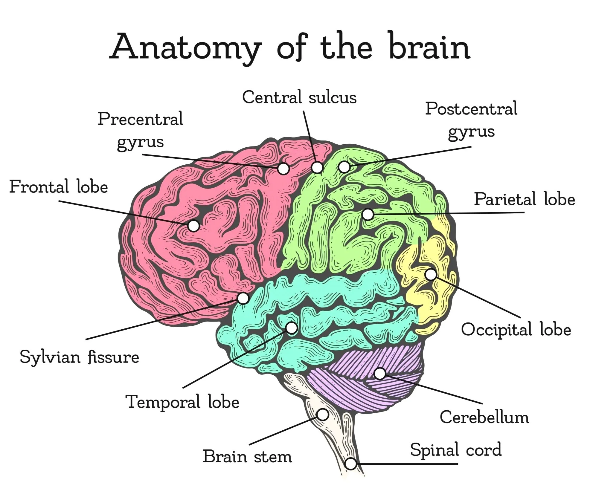
WHAT HAS BEEN LOCALISED IN THE NEOCORTEX
Click here to see an interactive brain map. PRESS
Now that we have examined what localisation of function means in the brain and discussed its importance, it is time to focus on the specific tasks that have been discovered and localised. These are primarily concerned with the following key areas:
MOTOR CENTRES
SOMATOSENSORY CENTRES
VISUAL CENTRES
AUDITORY CENTRES
LANGUAGE CENTRES: BROCA’S AND WERNICKE’S AREAS
Each of these areas plays a vital role in higher cognitive and sensory functions, and their localisation helps us understand the brain's structure and functioning in more detail.